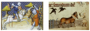
In our search for Medieval warhorses, we naturally will encounter a large quantity of horses not used in combat. The zooarchaeological record in Britain is full of horses, from the impressive Iron Age human/horse chariot burials, to the Post-Medieval remains of work horses. The task for those of us working on the bones of these animals is identifying signs that could indicate what the horses were being used for while alive. Because of the biased nature of preservation of biological remains, we frequently encounter bones and teeth, but rarely find evidence of more delicate materials, such as skin, fur, hair and muscle. Thus, it is our job to find proxies for these things with the faunal data we do have.
Muscles are a hugely important factor in how a horse moves, and those used for work (draught horses) would have utilised their muscles in very different ways from those used primarily for riding, or combat. Though we cannot study the muscles themselves, bones leave evidence of the nature of musculoskeletal loadings at the point where the muscle is attached to the bone. By looking at these areas of muscle attachment, we can investigate changes in robusticity, and changes in size, shape and surface complexity of horse bones.
As an analogy, imagine a draught horse as a front wheel drive car, deriving its power from the forelimbs, pulling with the aid of a harness. In contrast, in combat a horse is a rear wheel drive car, and would need to be able to perform short bursts of speed, and turn in tight, quick movements. It is likely this power would be derived from the hindlimbs. These vastly different movements repeated over the life of the animal would induce stress to the muscles, and the attached bones.
But, what can we expect to see on these bones? There are a few different ways that we are investigating these morphological changes, and a previous blog post discussed the 3D methods we are using. However, in-life movement and stress not only affect the surface of the bone, but also the internal structures. Studies of living horses have successfully used CT scanning and x-ray to investigate the internal structures of the lower limb bones (metapodia) providing a measurement of cortical thickness which has been shown to change depending on in-life usage of the horse. A few weeks ago, we used the x-ray in the archaeological science labs at the University of Exeter, to investigate the ability of x-ray to show the cortical thickness in archaeological bones.
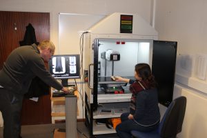
We investigated how the cortical thickness changed at different locations on the bone (i.e. near the ends and towards the centre of the shaft). This was preliminary investigative work to determine a sampling strategy for all the bones in our assemblage. We wanted to see how the cortical thickness changed along the bone, and decide on a standardised place to measure its thickness from. From the x-ray images of the horse bones, we will be able to measure the thickness of the cortical wall and compare this with the thickness of modern horses with known in-life activities.